
Simple Microscope Diagram (Parts labelled), Principle, Formula and Uses
This activity has been designed for use in homes and schools. Each microscope layout (both blank and the version with answers) are available as PDF downloads. You can view a more in-depth review of each part of the microscope here. Download the Label the Parts of the Microscope PDF printable version here.
Monday September 25 Parts of a Compound Light Microscope
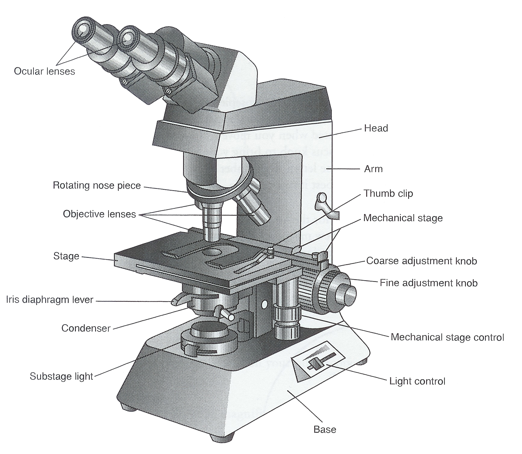
Parts Of a microscope. The main parts of a microscope that are easy to identify include: Head: The upper part of the microscope that houses the optical elements of the unit.; Base: The base is attached to a frame (arm) that is connected to the head of the device.The base of the microscope provides stability to the device and allows the user's hands to be free to manipulate other aspects of.

Parts of a Microscope The Comprehensive Guide Microscope and Laboratory Equipment Reviews
A Study of the Microscope and its Functions With a Labeled Diagram - Science Struck A Study of the Microscope and its Functions With a Labeled Diagram To better understand the structure and function of a microscope, we need to take a look at the labeled microscope diagrams of the compound and electron microscope.
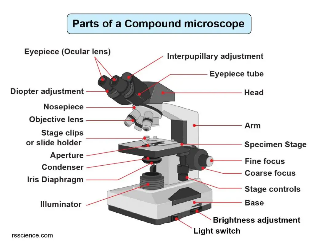
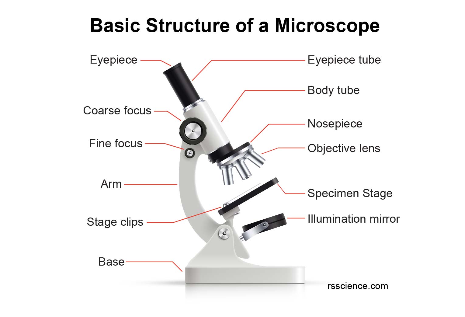
Compound Microscope Parts Labeled Diagram and their Functions Rs' Science
Explore the different parts of a microscope using a diagram, including the microscope lens, eyepiece, and stage. Updated: 10/13/2022 Create an account
Clipart microscope parts labeled WikiClipArt
Microscope Types (with labeled diagrams) and Functions Home / Microscope Types / Microscope - Types, Diagrams and Functions By Editorial Board October 13, 2022 Microscope - Let's split the name into two parts to understand what it actually means.

5 Types of Microscopes with Definitions, Principle, Uses, Labeled Diagrams
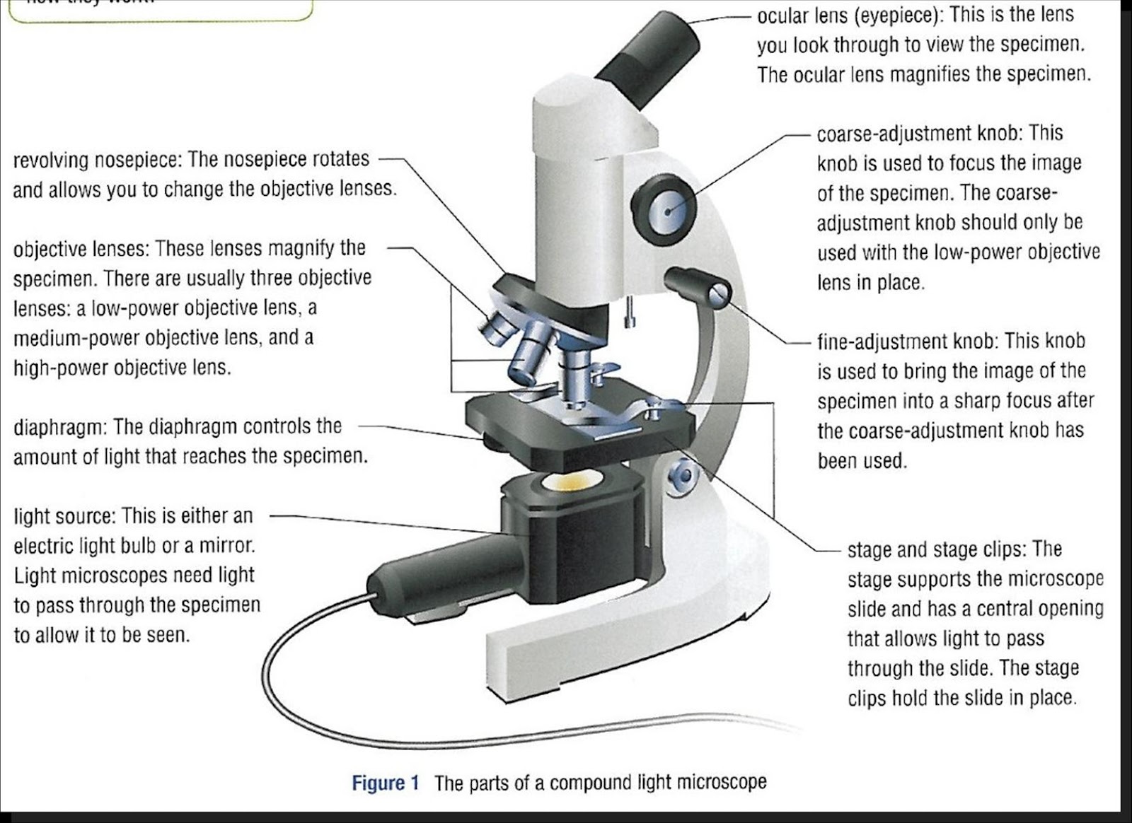
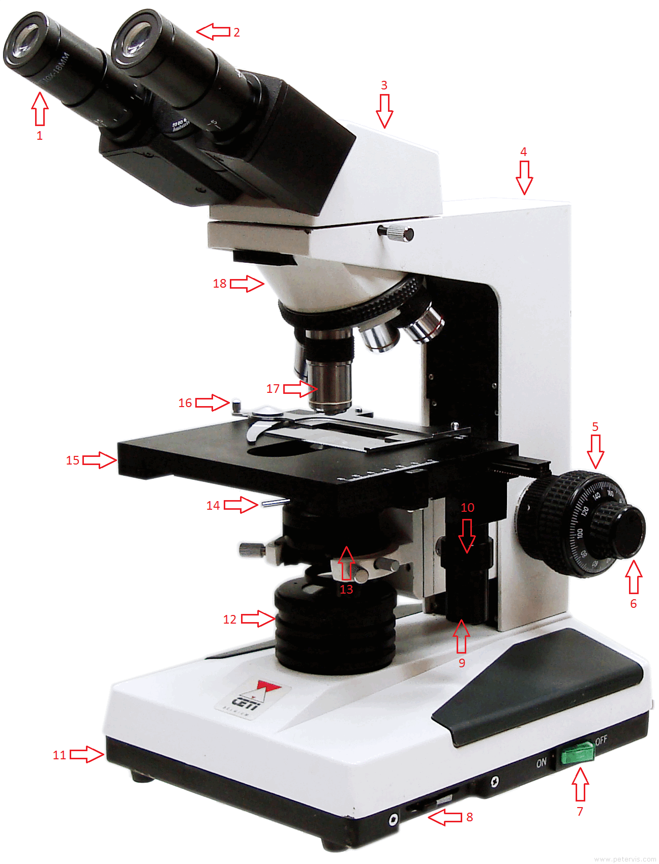
With Labeled Diagram and Functions How does a Compound Microscope Work? Before exploring microscope parts and functions, you should probably understand that the compound light microscope is more complicated than just a microscope with more than one lens.
Parts Parts And Functions Of A Microscope
Label the microscope Interactive Add to collection Use this interactive to identify and label the main parts of a microscope. Drag and drop the text labels onto the microscope diagram. eye piece lens coarse focus adjustment base stage diaphragm or iris high-power objective light source fine focus adjustment Download Exercise Tweet
What is a Microscope? Function and Magnification Rs' Science
The hand magnifying glass can magnify about 3 to 20×. Single-lensed simple microscopes can magnify up to 300×—and are capable of revealing bacteria —while compound microscopes can magnify up to 2,000×. A simple microscope can resolve below 1 micrometre (μm; one millionth of a metre); a compound microscope can resolve down to about 0.2 μm.
Labelled Diagram of Microscope Parts
Table of Contents History of Microscopy: Overview What is a light microscope? Figure: Diagram of Light Microscopes, created with biorender.com Principle of a light microscope (optical microscope) Types of light microscopes (optical microscope) Brightfield Light Microscope (Compound light microscope)
Parts of a Microscope Labeling Activity
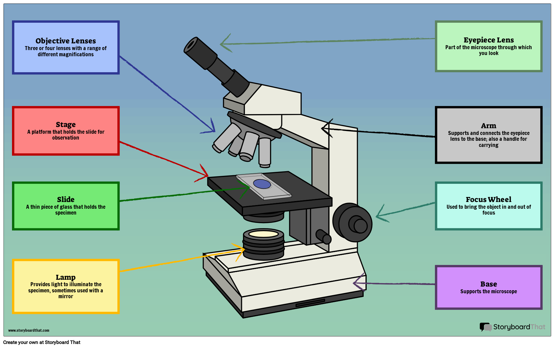
Diaphragm (Iris) Condenser Aperture Stage Objective lens Body Tube Ocular Lens (eye-piece) Coarse and Fine Adjustment Knob Arm Base Microscope Worksheet The Light Microscope Light microscopes are used to examine cells at relatively low magnifications. Magnifications of about 2000X are the upper limit for light microscopes.
How to Use a Microscope
The working principle of a simple microscope is that when a lens is held close to the eye, a virtual, magnified and erect image of a specimen is formed at the least possible distance from which a human eye can discern objects clearly. Magnification formula The magnification power of a simple microscope is expressed as: M = 1 + D/F Where

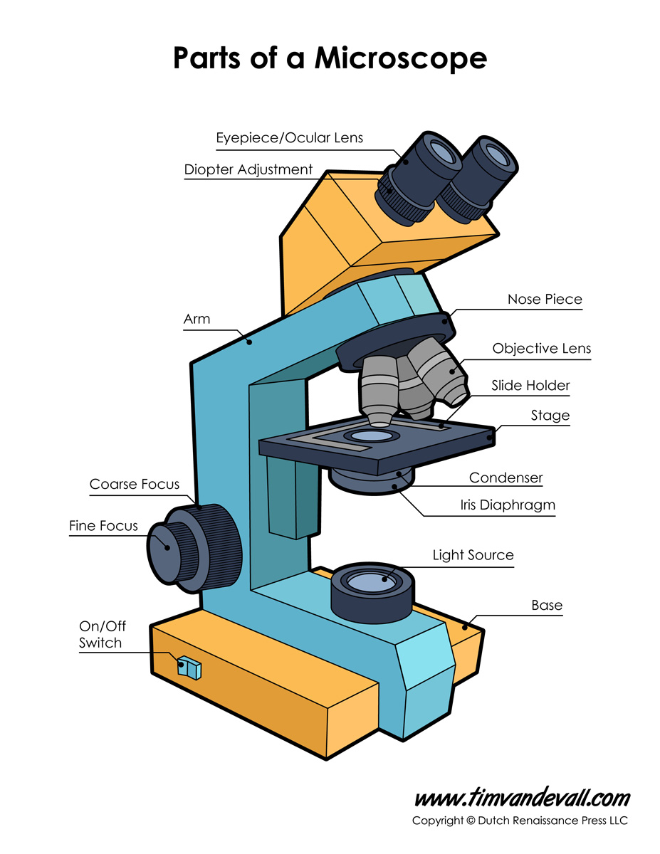
Parts of a microscope with functions and labeled diagram
There are three major structural parts of a compound microscope. The head includes the upper part of the microscope, which houses the most critical optical components, and the eyepiece tube of the microscope. The base acts as the foundation of microscopes and houses the illuminator. The arm connects between the base and the head parts.
Ag Biology Unit 2
A labeled diagram of microscope parts furnishes comprehensive information regarding their composition and spatial arrangement within the microscope, enabling researchers to comprehend their function effectively. In this comprehensive article, we will delve into the intricate parts of the microscope, exploring their functions in detail.
16 Parts of a Compound Microscope Diagrams and Video Microscope Clarity
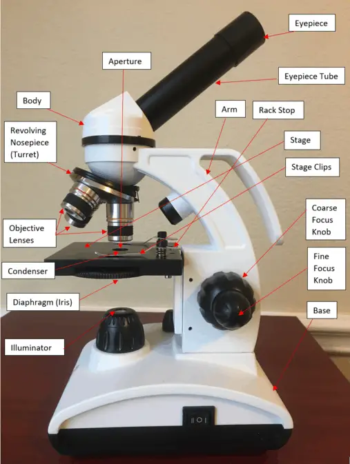
Parts of the Microscope with Labeling (also Free Printouts) A microscope is one of the invaluable tools in the laboratory setting. It is used to observe things that cannot be seen by the naked eye. Table of Contents 1. Eyepiece 2. Body tube/Head 3. Turret/Nose piece 4. Objective lenses 5. Knobs (fine and coarse) 6. Stage and stage clips 7. Aperture
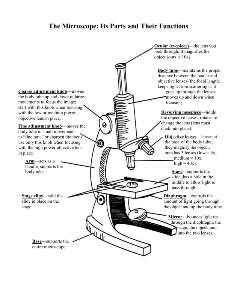
The Microscope Its Parts and Their Functions
The web page titled "Parts of a Microscope with Labeled Diagram and Functions" has the following key takeaways: 🔍 The microscope is an essential tool for scientists, researchers, and medical professionals. 🧬 The main function of a microscope is to provide a magnified view of small objects or organisms, such as bacteria, cells, or tissues.
1.5 Microscopy Biology LibreTexts
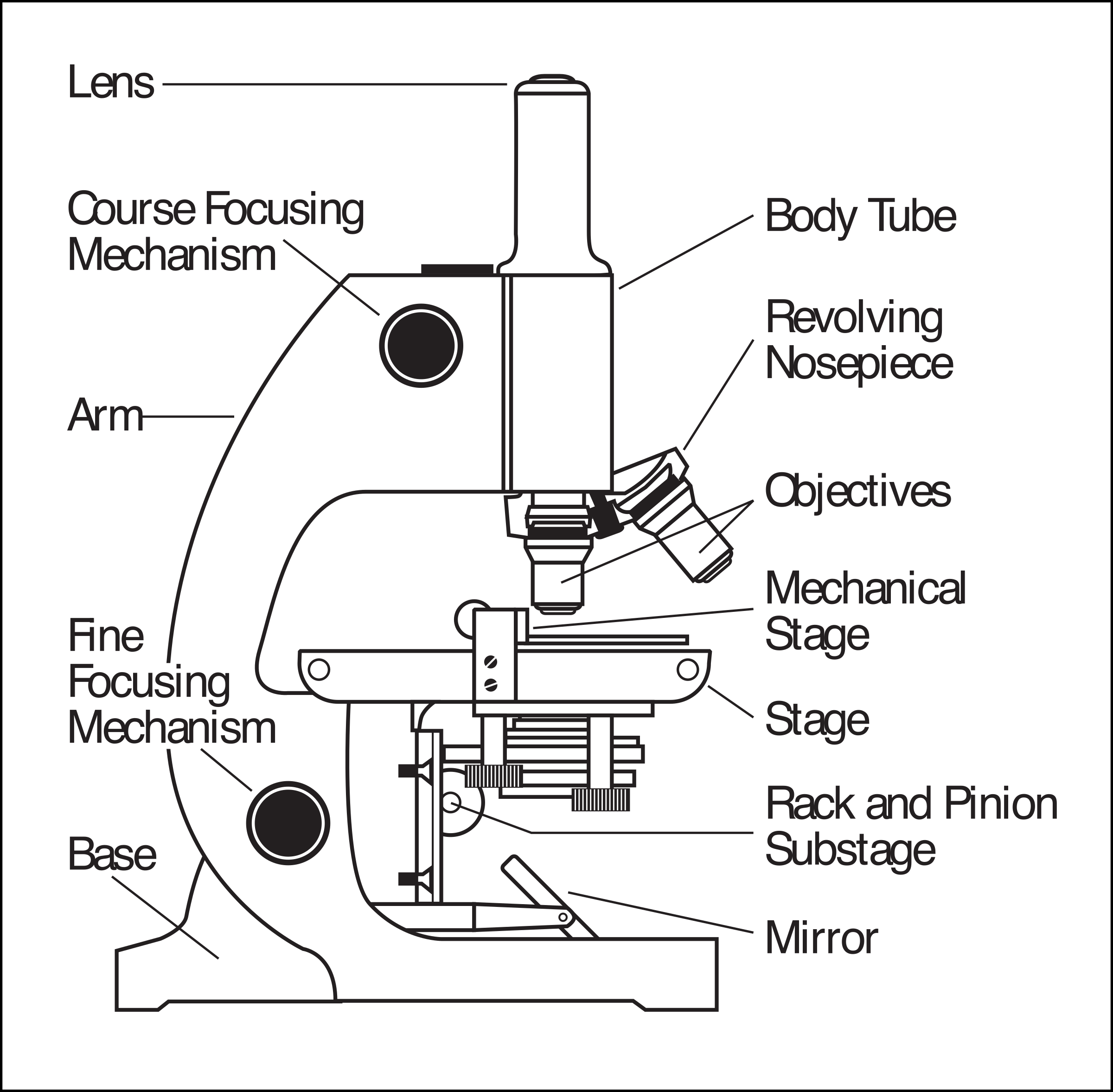
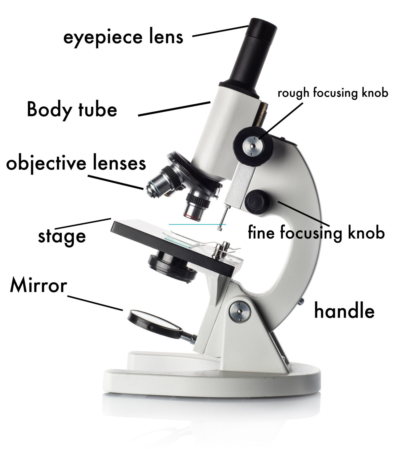
The microscope illustrated in Figure 5 below was manufactured by Hugh Powell and Peter Lealand around 1850. The tripod base provided a sturdy support for the microscope, which many people consider the most advanced of its period. Parts of a Powell and Leland Microscope Diagram